Resources
We have a variety of CT-related resources available.
Patient CT projection data library
The CT Clinical Innovation Center has developed and made publicly available data from hundreds of patient CT scans, including exams of the head, chest and abdomen from Siemens scanners and GE scanners. This information, which is available through The Cancer Imaging Archive (TCIA), is called the Low-Dose CT Image and Projection Data collection.
The library has data from 299 CT exams of the head, chest and abdomen — 150 from Siemens scanners and 149 from GE scanners. All exams have routine-dose projection data and simulated low-dose projection data, full-dose images, and clinical data identifying all pathology.
This library is unique because for the first time ever it includes the X-ray projection data used to create cross-sectional images. These data are critical for developing advanced reconstruction and denoising techniques, including those using artificial intelligence.
Related publications
Moen TR, Chen B, Holmes III DR, Duan X, Yu Z, Yu L, Leng S, Fletcher JG and McCollough CH. Low‐dose CT image and projection dataset. Medical Physics. 2021;48:902.
McCollough C, Chen B, Holmes III D R, Duan X, Yu Z, Yu L, Leng S and Fletcher J. (2020). LDCT-and-Projection-data: Low Dose CT Image and Projection Data (Version 6) [Data set]. The Cancer Imaging Archive. 2020.
Chen B, Duan X, Yu Z, Leng S, Yu L and McCollough CH. Technical Note: Development and validation of an open data format for CT projection data. Medical Physics. 2015;42:6964.
Yu L, Shiung MM, Jondal D and McCollough CH. Development and validation of a practical lower-dose-simulation tool for optimizing computed tomography scan protocols. Journal of Computer Assisted Tomography. 2012;36:477.
CT Grand Challenge data
A library of CT image and projection data is available for use in developing and evaluating CT reconstruction and denoising techniques.
These data are from the 2016 international Low Dose CT Grand Challenge. Participants from more than 20 countries were challenged to quantitatively assess the diagnostic performance of denoising and iterative reconstruction techniques on common low-dose patient CT datasets using a detection task, allowing the direct comparison of various algorithms. The results provided an indication of the range of performances achieved using different classes of denoising and iterative reconstruction techniques.
Our CT scanners
Since 2004, the CT Clinical Innovation Center has operated eight different first-of-its-kind CT systems. Our current CT system is a Siemens NAEOTOM Alpha. The Alpha is a dual-source photon-counting detector (PCD) CT scanner. It is the world's first commercially available clinical PCD-CT system.
Our journey with PCD-CT technology began in 2014, when the first PCD prototype, the Siemens SOMATOM CounT, was installed at Mayo Clinic as an investigational scanner. In 2020, the second-generation prototype PCD system, the SOMATOM Count Plus, was installed to continue investigating clinical photon-counting CT and assessing image quality with the technology improvements developed since the first-generation system was built.
The NAEOTOM Alpha was installed in April 2021 as an investigational prototype system. It became an official clinical CT system after clearance from the U.S. Food and Drug Administration in September 2021.
Because this is an emerging technology and the first-of-its-kind scanner, we have numerous opportunities to explore and identify advantages and new applications for CT that can improve diagnostic imaging and care.
Phantoms and dosimeters
To facilitate our research studies, we have a large inventory of test objects, called phantoms, and devices to measure radiation dose, called dosimeters.
Phantoms are particularly helpful in gathering data that simulate the data collected from people undergoing CT scanning. For example, if we want to study how reducing the radiation dose can affect the image quality, we use a test phantom and take scans at different dose levels to compare image quality. Phantoms enable us to acquire scans in which the resulting images look very much like real people, allowing us to conduct many research experiments without exposing people to unnecessary radiation.
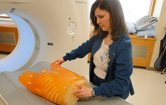
A CT technologist places a phantom designed to look like a human chest on the bed of a CT scanner.
Software
Specialized software we have developed includes:
We also have software tools to read and modify projection data from Siemens scanners and to return the modified data to the scanner for image reconstruction. The ability to read, modify and return projection data has allowed us to develop advanced postprocessing tools for projection data modification, such as metal artifact reduction. In turn, this allows us to help patients by improving the quality of their CT images.