 Mapping atypical pain processing
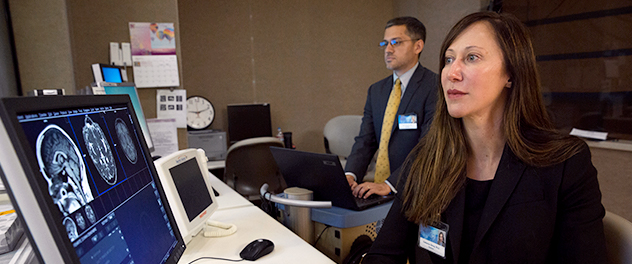
Using brain imaging techniques to analyze and identify imaging biomarkers for concussion, migraine and other headache disorders, Dr. Schwedt and his team study how alterations in brain structure or function relate to pain processing.
Overview
The Neuroimaging of Headache Disorders Laboratory of Todd J. Schwedt, M.D., and Cat Chong, Ph.D., at Mayo Clinic's campus in Phoenix, Arizona, uses magnetic resonance imaging (MRI) and other types of imaging to study migraine, post-traumatic headache and other headaches. Correlating patient symptoms with brain imaging helps the team better understand the underlying causes of migraine and headache disorders.
More specifically, the aim is to pinpoint the areas of the brain associated with pain and other symptoms and investigate the neuroimaging commonalities and differences among headache disorders. The laboratory uses computational modeling techniques that enable the classification of specific headache disorders and prediction of patient outcomes based on structural and functional brain MRI data.