-

Determining disease severity
To help determine the severity of myocarditis and dilated cardiomyopathy, a lab staff member is using histology staining techniques such as haematoxylon and eosin to identify inflammation, trichrome blue and picrosirius red stain to examine fibrosis, and toluidine blue to detect mast cell degranulation.
-
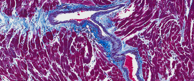
Understanding fibrosis
We study why fibrosis is laid down in the heart after myocarditis in order to develop new prevention strategies. Fibrosis can be laid down on the pericardium, in the myocardium or around the vessels, as shown here in blue.
-
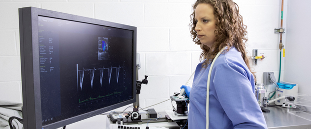
Evaluating heart function
Echocardiogram is used to determine heart function in our myocarditis and dilated cardiomyopathy model using equipment that's similar to the equipment used for patients. We're able to assess how well the heart is pumping and how it looks structurally and to detect heart failure. Here, former lab staff member Katelyn A. Bruno, Ph.D., examines diastolic heart function in myocarditis to determine the effect of a novel potential regenerative medicine therapy.
-
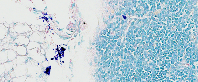
Mast cell degranulation in the heart
Mast cells are key immune cells in many conditions, including allergies and asthma. We have found that mast cell degranulation, as seen in this image, also is important for disease progression in myocarditis.
-

Microscopy
Histological analysis is a key component when studying myocarditis.
-
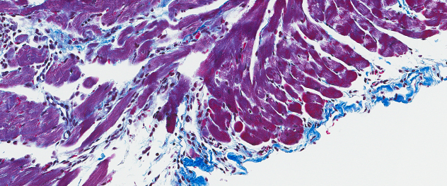
Fibrosis along the pericardium
Acute myocarditis can cause scar tissue to be laid down, as seen in this image of fibrosis along the pericardium, leading to chronic heart failure and possibly the need for a heart transplant.
-
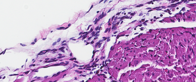
Pericardial inflammation
The first step in every experiment we conduct is determining how much inflammation is in the heart. When the inflammation is along the outside of the heart as indicated here, it's known as pericarditis.
-

Immune cell populations
Assessment of specific immune cell populations as seen in this image can help determine the mechanism of disease.
-
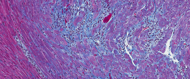
Chronic myocarditis with fibrosis
In some people, myocarditis can lead to chronic myocarditis and heart failure. Immune cells, shown here as round dark blue cells, and fibrosis, seen as areas of bright blue, infiltrate the heart, worsening heart function.
Research
As an expert in myocarditis, dilated cardiomyopathy and heart failure, Dr. Fairweather leads a research team seeking advances in diagnostic techniques and novel therapies. Translational research and clinical sample analysis are key for breakthroughs to occur in the field.
Areas of research
Our lab engages in five main areas of research:
- Identifying immunomechanisms contributing to myocarditis, dilated cardiomyopathy and heart failure.
- Understanding how sex differences in inflammation contribute to the pathogenesis of cardiovascular and autoimmune conditions.
- Identifying biomarkers that improve the diagnosis of cardiac conditions and other chronic inflammatory conditions.
- Studying how environmental factors such as infection, vitamin D levels and chemicals can cause chronic inflammation.
- Understanding how regenerative medicine therapies prevent chronic conditions.
Collaborators
Our lab collaborates with other researchers from Mayo Clinic whose expertise in cardiovascular disease, multiple myeloma, chronic lymphocytic leukemia, myocarditis and individualized medicine are helping us make advances in diagnosis and treatment.
We collaborate with these Mayo Clinic researchers:
- Atta Behfar, M.D., Ph.D.
- Asher A. Chanan-Khan, M.B.B.S., M.D.
- Leslie T. Cooper Jr., M.D.
- Jonathan B. Hoyne, Ph.D.
- Khashayarsha Khazaie, Ph.D.
- Carolyn Landolfo, M.D.
- Debabrata (Dev) Mukhopadhyay, Ph.D.
- Todd D. Rozen, M.D.
- Lynsey A. Seim, M.D., M.B.A.
- Brian P. Shapiro, M.D., M.A.
- Shane A. Shapiro, M.D.
- Peter Storz, Ph.D.